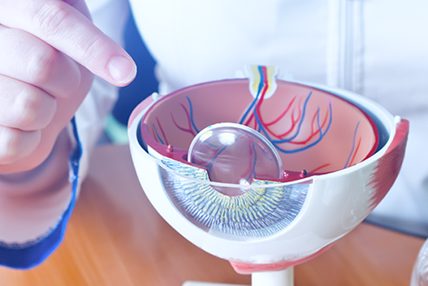
Ophthalmology
Clear Vision, Healthy Eyes: The Vital Role of Routine Eye Exams in Early Detection and Prevention
May 2026
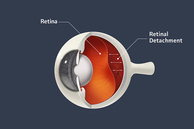
The retina, a delicate layer of tissue lining the back of the eye, plays a crucial role in vision. However, when the retina peels away from its normal position, a condition known as retinal detachment occurs, demanding immediate attention. In this article, we will explore the urgency of addressing retinal detachment, the signs and symptoms, and the surgical interventions that can help restore vision and prevent permanent vision loss.
Understanding Retinal Detachment: A Vision-Threatening Emergency :
Retinal detachment occurs when the retina, responsible for capturing visual images and sending them to the brain, pulls away from the underlying supportive tissue. This can happen suddenly, and the condition is considered a medical emergency that requires prompt intervention.
Risk Factors :
Recognizing the Signs and Symptoms: Act Swiftly, Save Sight
Early detection is crucial in preventing permanent vision loss associated with retinal detachment. Recognizing the signs and symptoms can prompt timely intervention. These may include :
Emergency Intervention: Seeking Immediate Medical Attention :
If you suspect retinal detachment, seeking emergency medical attention is imperative. Delay in treatment can lead to severe vision impairment or even permanent blindness. An ophthalmologist will conduct a thorough examination, including a dilated eye exam, to diagnose the detachment.
Surgical Interventions: Restoring the Retina for Clear Vision :
Post-Surgery Care and Recovery: Nurturing the Healing Process :
Following retinal detachment surgery, patients will require diligent post-operative care, including the use of prescribed eye drops, limitations on physical activities, and routine follow-up appointments. The success of the surgical intervention depends on factors such as the extent of detachment and the promptness of treatment.
Retinal detachment is a sight-threatening emergency that demands immediate attention and surgical intervention. Recognizing the signs, seeking prompt medical help, and undergoing appropriate surgical procedures can significantly increase the chances of preserving vision and preventing irreversible damage. Regular eye check-ups and awareness of risk factors can contribute to early detection, highlighting the importance of maintaining eye health and preventing complications associated with retinal detachment.